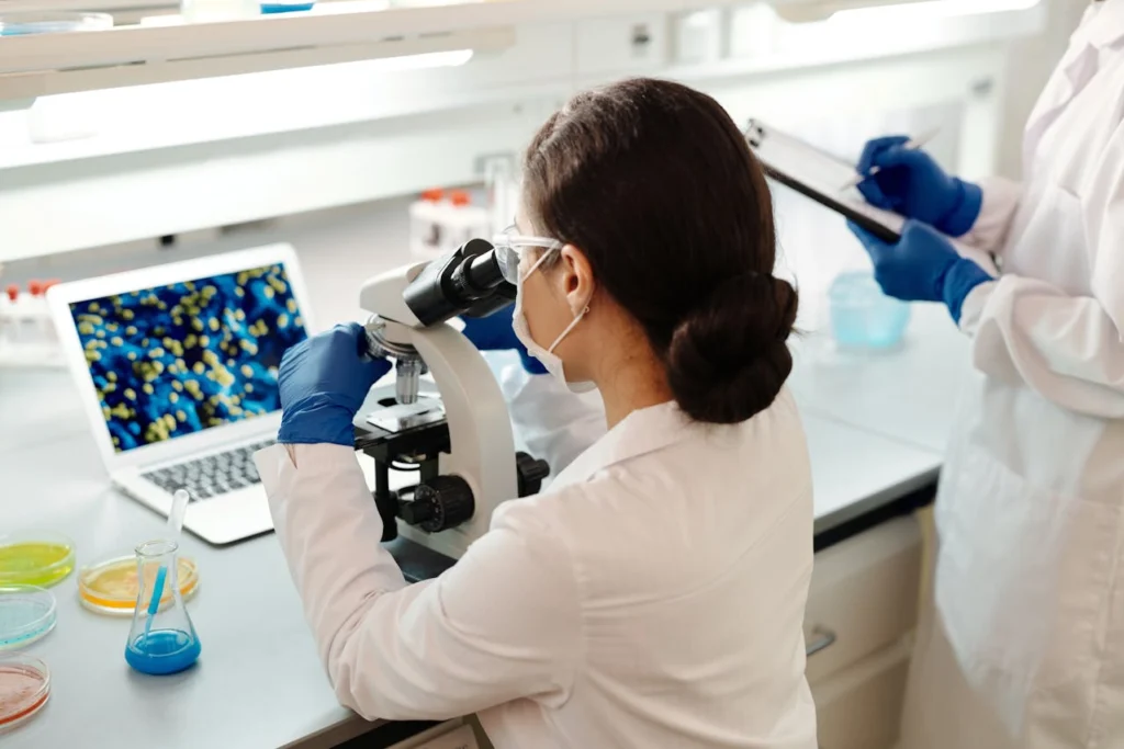
Fluorescence microscopy shows us a world of light and color. It reveals the hidden architecture of cells. But that raw image is just the beginning. It is often blurry. It can be noisy. The real magic happens after the photo is taken.
This is where image processing software enters the story. It transforms a raw snapshot into robust, quantitative data. This step is not just about making pictures look nice. It is about making them scientifically true. The journey from a raw capture to a reliable result is fascinating. Let’s dive in.
Clarifying the Foundation
First, let’s set the stage. Many people wonder what is fluorescence microscope technology actually doing? In simple terms, it uses specific wavelengths of light. This light causes molecules in a sample to glow. Scientists tag different cellular structures with these glowing markers. The microscope captures this emission.
The result is a detailed map of cellular activity. But this map is rarely perfect straight from the instrument. It needs development, much like a photograph from an old film camera. The camera sensor itself introduces artifacts. The optics have physical limitations. These factors mean we never get a perfect representation right away.
Taming the Digital Noise
Every image has noise. This appears as a grainy, speckled texture. It comes from the camera sensor itself. It can also come from stray light. This noise obscures the real signal. It makes structures look fuzzy and ill-defined. Processing software uses clever algorithms to fix this.
Filters like Gaussian blur can smooth out the background. More advanced techniques exist too. They intelligently separate the true signal from this random noise. The result is a much cleaner image. The actual biological information shines through without distraction. This is the first and most critical step toward clarity.
Bringing Blur Into Sharp Focus
Blur is another major enemy. It comes from many sources. The lens itself can cause it. So can the movement of living samples. Out-of-focus light from above and below the focal plane also contributes. This haze reduces contrast and hides detail.
Deconvolution is a powerful software solution. It mathematically reassigns this misplaced light. It uses a model of the microscope’s specific blur pattern. The effect is remarkable. Hazy blobs become sharp, crisp structures. You can see details you never knew were there. This process literally adds resolution back into your data.
Making Measurements Meaningful
Science needs numbers. Simply looking at an image is not enough. Researchers need to quantify everything. They measure protein levels. They track the size and shape of organelles. They count individual molecules. Processing software provides these tools.
It can automatically outline cells. It can measure fluorescence intensity pixel by pixel. It can even track a moving particle over time. This turns a subjective observation into an objective, numerical fact. This is the heart of accurate data analysis. Without quantification, an image is just a picture. With it, the image becomes evidence.
Isolating the Specific Signal
Real samples are messy. You might be looking at one protein. But other things in the cell glow too. This is called background or autofluorescence. It creates a false signal. Software helps subtract this unwanted glow.
You define a region that should be dark. The software calculates the background level in that area. It then subtracts that value from the entire image. What remains is the true, specific signal from your target. This correction is vital for any kind of accurate intensity measurement. It ensures you are measuring your target, not the cellular junk.
Creating a Clear Visual Story
Sometimes, you need to present your data. A journal cover demands a striking visual. A conference presentation needs clear graphics. Processing software is essential here. It allows for the adjustment of brightness and contrast. This makes features more visible. It does so without altering the underlying quantitative data.
You can also merge channels. This creates those classic red-and-green merge images that show protein co-localization. A well-processed image tells a compelling and truthful story. It communicates your hard work effectively to others.
The Non-Negotiable Workflow
In the end, processing is not optional. It is a fundamental part of modern microscopy. A raw image is just a collection of pixel values. It is full of optical imperfections and physical artifacts. Software corrects these flaws. It unveils the true biological information. It transforms beautiful pictures into hard, trustworthy data.
This partnership between the microscope and the computer is what drives discovery forward. It ensures that what we see is what we actually get. Your data is only as strong as your processing pipeline. Ignore this step at your own peril.
- About the Author
- Latest Posts
Our Editorial Team are writers and experts in their field. Their views and opinions may not always be the views of Wellbeing Magazine. If you are under the direction of medical supervision please speak to your doctor or therapist before following the advice and recommendations in these articles.