For millions of older adults worldwide, life is often tarnished by the persistent, gnawing presence of chronic pain management. Among the most prevalent causes of this discomfort is osteoarthritis, a condition frequently dismissed by patients as an inevitable part of aging. However, a significant barrier to effective treatment is not just the physical ailment itself, but a profound lack of understanding. When a physician hands a senior patient a paper filled with dense clinical text or shows them a static, grayscale X-ray, the comprehension is hard to achieve. To a layperson, “joint space narrowing” or “osteophyte formation” are abstract concepts.
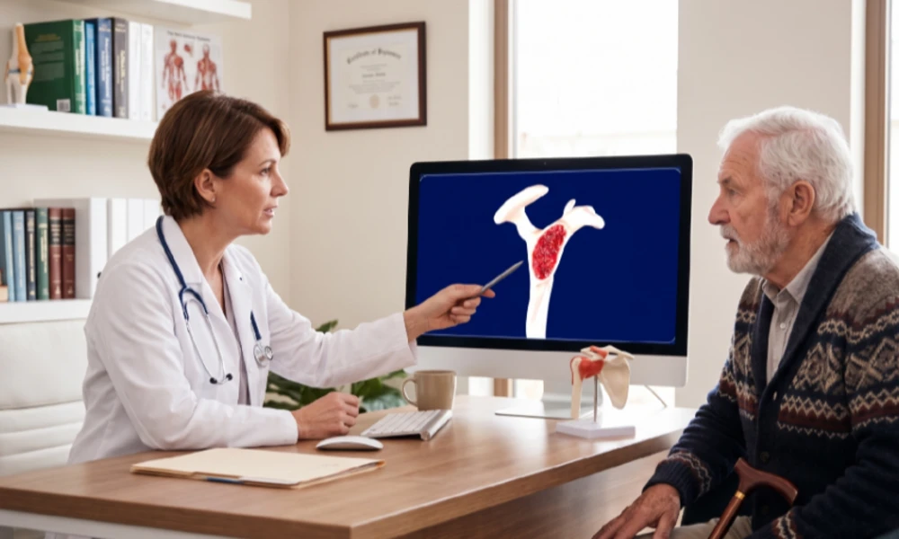
The use of high-fidelity 3D medical animation is changing this narrative. By transforming invisible, internal biological processes into clear, cinematic stories, technology is providing a path toward patient empowerment and improved education.
The communication gap in senior healthcare
The psychological impact of chronic pain is often followed by confusion. Many seniors suffering from joint degeneration struggle to follow complex treatment regimens because they do not fundamentally grasp what is happening inside their bodies. When this understanding is missing, the necessity of treatment, such as physical therapy, medication, or lifestyle changes, is often ignored.
Textual diagnoses are limited, as they describe a state of being but fail to illustrate a process. For a patient experiencing a decline in senior mobility, hearing that their “cartilage is wearing thin” does not provide the same impact as seeing the biomechanics of pain in motion. This is where a mechanism of disease animation steps in to bridge the gap between clinical expertise and patient comprehension.
Peering beneath the surface: the mechanism of disease
The true power of animation lies in its ability to take the viewer where no camera can go. Through detailed 3D visualization, patients can witness the microscopic reality of their condition.
In the context of arthritis, an animation can start with the smooth, glistening surface of a healthy synovial joint. As the animation progresses, it reveals the gradual thinning of the articular cartilage, the increased friction between bone surfaces, and the subsequent inflammatory response. Seeing the “fraying” of the tissue at a cellular level makes the abstract concept of “wear and tear” visceral and real.
By visualizing the biomechanics of pain, the patient can see exactly why a certain movement hurts. They can see how bone-on-bone contact triggers nerve signals, providing a logical framework for their symptoms. This clarity often reduces the fear and anxiety associated with the unknown, allowing the patient to view their condition as a mechanical challenge that can be managed.
Empowering patients and caregivers
Understanding is the first step toward compliance. When a patient understands that their exercises are designed to strengthen the muscles supporting a degenerated joint, they are more likely to perform them. This is particularly crucial in in-home care settings, where the absence of a constant medical supervisor means the patient must be self-motivated.
3D animation serves as a tool for patient empowerment by:
- Validating the patient’s experience. Seeing the inflammation and structural damage on screen confirms that their pain is real and has a physical source.
- Clarifying treatment goals. If a patient sees how a specific injection or supplement acts as a lubricant or anti-inflammatory within the joint space, they perceive the value of the intervention more clearly.
- Encouraging proactive management. Knowledge of how joint degeneration progresses can motivate seniors to adopt ergonomic changes early, preserving their independence for longer.
Enhancing senior mobility and quality of life
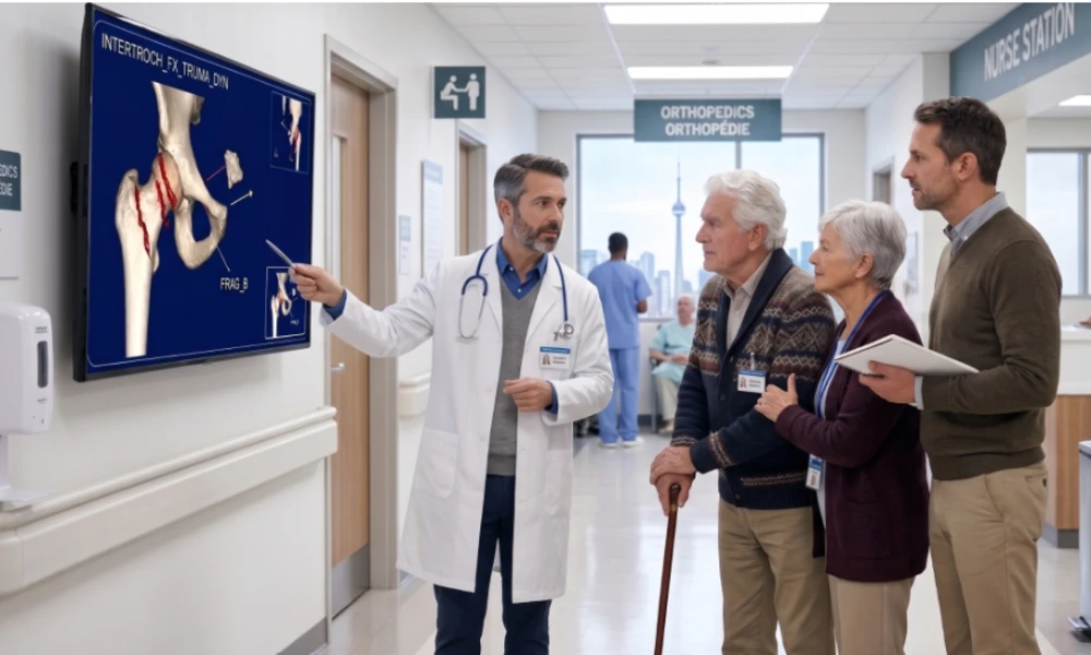
The ultimate goal of visualizing arthritis is to facilitate arthritis relief and restore senior mobility. As the global population ages, the burden on healthcare systems increases. Tools that can be used remotely on tablets or during telehealth consultations to explain musculoskeletal health are becoming indispensable.
For family members providing in-home care, these animations are equally valuable. They allow them to understand the physical limitations of their relatives, fostering empathy and helping them assist with mobility exercises more effectively. Instead of a source of frustration, the “stiff joint” becomes a clearly understood biological reality that requires specific, gentle handling.
Companies like VOKA are leading this visual revolution. By producing realistic 3D models that accurately reflect the latest medical research, they provide clinicians with a visual “language” that overcomes age and education levels. Whether it is a surgeon explaining a total hip replacement or a general practitioner discussing osteoarthritis management, these animations ensure that the patient is an informed partner in their own care.
Conclusion
Demystifying the biomechanics of pain is essential for the future of geriatric care. As we move away from paternalistic medicine toward a collaborative model, the patient’s understanding becomes more and more vital. 3D medical animation doesn’t just show the destruction of cartilage but makes the invisible visible, providing seniors not just with a diagnosis, but with the clarity required for long-term chronic pain management and a higher quality of life.
- About the Author
- Latest Posts
Our Editorial Team are writers and experts in their field. Their views and opinions may not always be the views of Wellbeing Magazine. If you are under the direction of medical supervision please speak to your doctor or therapist before following the advice and recommendations in these articles.